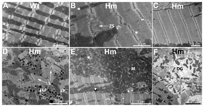
Figure 3. Electron microscopy of wild type and homozygous mutant left ventricle from rats one year of age.
Samples were fixed, embedded, sectioned, and stained using standard methods prior to electron microscopic examination (see Materials and Methods for full details). A. Typical wild type cardiomyocyte appearance with myofibrils flanked by mitochondria. B. Homozygote with evidence of Z line streaming (arrow, ZS). C. Homozygote with exceptionally wide myofibril region. D. Homozygote with dark stained lipofuscin granules (arrow, LF). Filament bundles were observed with different orientations in the XY plane (arrows, L) and also running perpendicularly (arrow, P) E. Homozygote with a large cluster of mitochondria (M). F. Homozygote cardiomyocyte region showing myofibril disintegration (DG).

