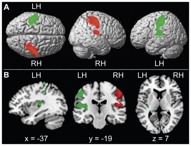
Figure 2.

A Activation maps for the comparisons target-absent AL > AR and target-absent AR > AL, thresholded at p < .001 uncorrected. Both contrasts revealed increased activity in the contralateral primary and secondary somatosensory cortices. Attention to the right hand resulted in additional activation in left posterior insula. In target-absent AL > AR, right SI is revealed (red) with the MNI coordinates of +54, -19, +49 (x, y, z) for the peak activation as well as right SII with peak activation at +48, -16, +13 (x, y, z). In target-absent AR > AL, left SI is revealed (green) with the MNI coordinates of -45, -28, +49 for the peak activation. Moreover, left SII is significantly activated with peak activation at -48,-22,+16 (x, y, z) as well as left posterior insula with peak activation is at -33, -19, +10 (x, y, z). The contrast maps have been superimposed on an SPM template. From left to right, dorsal and lateral views of right and left hemisphere are depicted. Abbreviation for the left and right hemisphere is LH and RH, respectively. B Activation maps for the contrast target-absent AL > AR in green and target-absent AR > AL in red, thresholded at p < .001, uncorrected. Activity in the left primary somatosensory cortex is depicted in the upper cluster of the first two pictures. In the lower cluster the activation over the left posterior insula and left secondary somatosensory cortex is shown. Activity in the posterior insula can also be seen in the third image. From left to right sagittal, coronal and horizontal slices are depicted. Abbreviation for the left and right hemisphere is LH and RH, respectively.
