
Figure 1. Relative quantification of alpha-cardiac actin 1 (ACTC1) by immunoblotting.
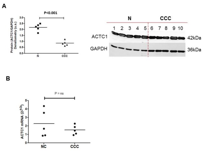
Myocardial samples were obtained from the left ventricular free wall of the hearts of patients with severe CCC and end-stage heart failure, at the time of heart transplantation. Samples from five hearts from CCC patients (at least two positive results in three independent anti-T. cruzi serology tests, as indicated above), and from healthy hearts from organ donors not used for transplantation for technical reasons were used. Immunoblotting and protein quantification were done in duplicate. A. The immunoblot and the protein quantification result of the first experiment are presented here. The central line represents the median. Representative results from two experiments are shown here. A Mann-Whitney test was performed and differences were considered significant if P<0.001.
B. Real-time quantitative PCR was carried out on the same samples. All the samples were tested in triplicate with GAPDH, the expression of which has been shown to vary little between human myocardial tissue samples. Data were normalized and the relative levels of each mRNA were calculated by the 2-ΔCt method.