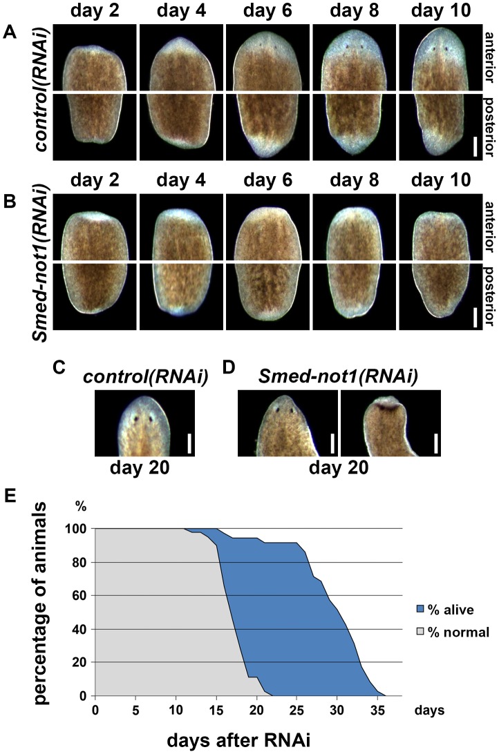
Figure 2. Smed-not1 is required for planarian regeneration and homeostatic cell turnover.
(A–B) Control(RNAi) (A) and Smed-not1(RNAi) (B) animals cut 5 days after RNAi, and monitored every 2 days after transection. Top panels: anterior wounds; bottom panels: posterior wounds. Smed-not1 animals are able to produce blastemas (white tissue) in both anterior and posterior wounds but fail to regenerate. (C–D) Intact control(RNAi) (C) and Smed-not1(RNAi) (D) animals 20 days after RNAi, anterior side. Smed-not1 animals 20 days after RNAi display variable levels of head regression defects. (E) Survival curve of Smed-not1 animals (N = 40), indicating the percentage of animals without any defects (grey) and the percentage alive (blue). All control(RNAi) animals survived without defects for more than 40 days (N = 40). Scale bars: 500 µm. See also Figure S3.

