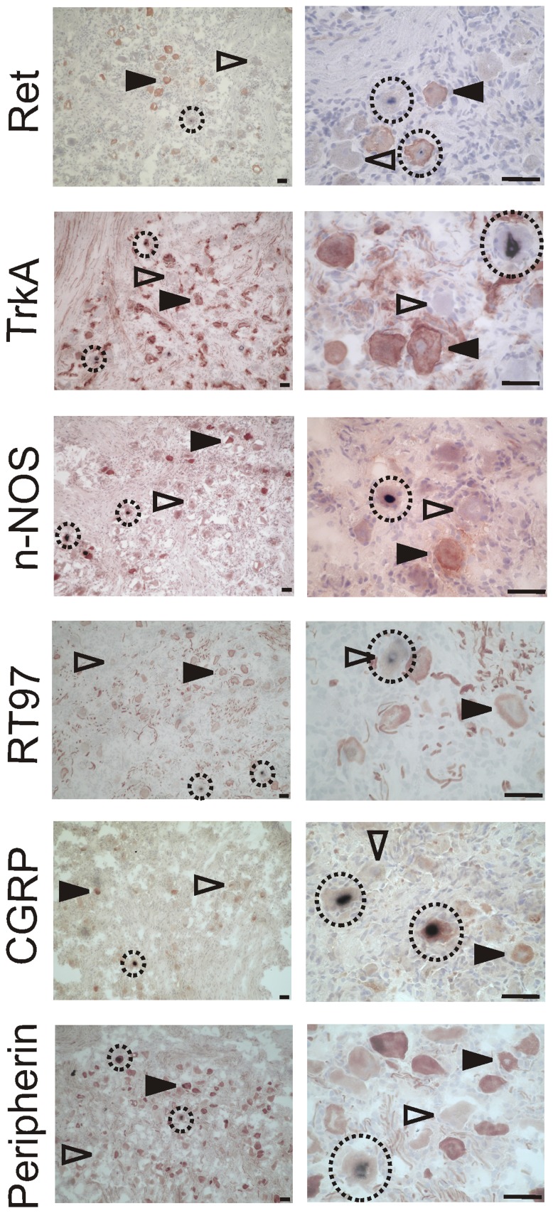
Figure 4. Detection of HSV-1 LAT in neurons expressing different maker proteins in the human TG.
The micrographs in the left column are taken at 100x magnification for an overview, those in the right column at 400x for a more detailed view. The name of the marker used is indicated in each row. Marker proteins are labeled with DAB (brown), LAT using NBT (purple), and the tissue was counterstained with haematoxylin. Open arrowheads indicate marker negative cells, closed arrowheads marker positive cells. LAT positive cells are circled. Scale bars represent 50 µm in all cases.

