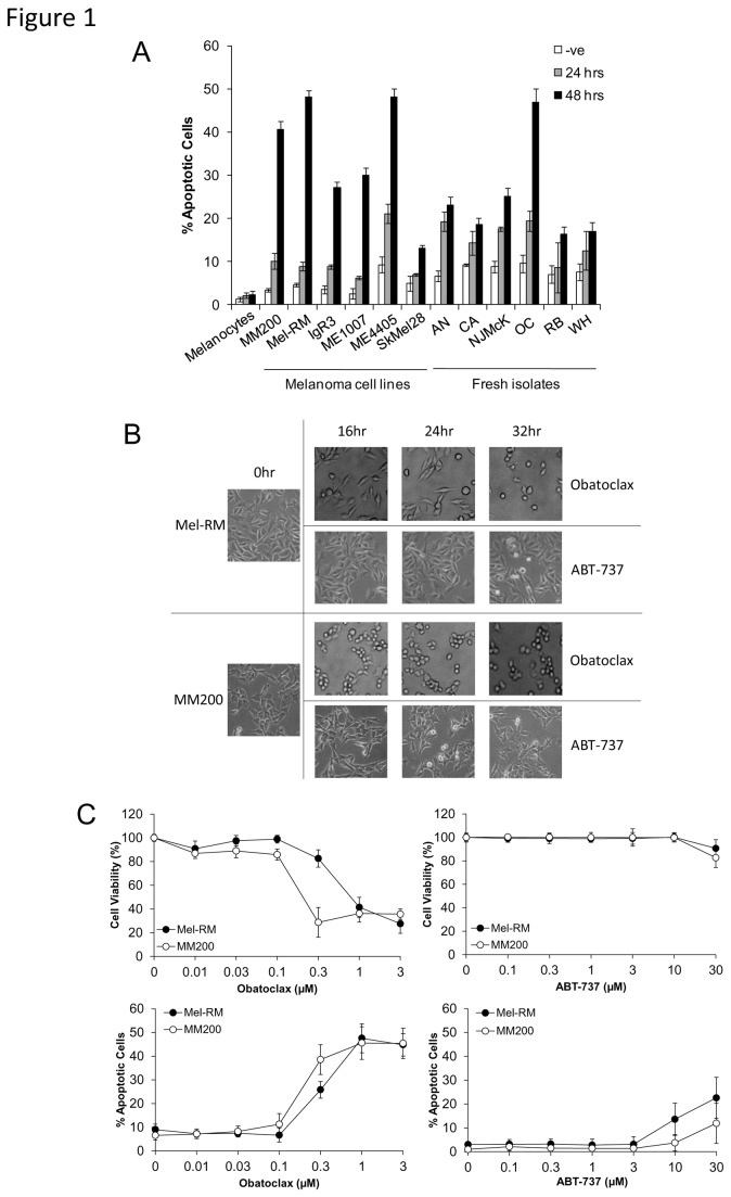
Figure 1. Obatoclax and ABT-737 induce apoptosis in melanoma cell lines.
A. Summary of studies on a panel of melanoma cells and fresh melanoma isolates with normal cells (melanocytes). Following treatment of cells with Obatoclax (1μM) for 24 and 48 hours, cells were subjected to measurement of apoptosis by the propidium iodide method. Points, mean of three individual experiments; bars, SE.
B. Treatment with Obatoclax, but not ABT-737, causes morphological changes in melanoma cells. Mel-RM and MM200 cells were treated with Obatoclax (1μM) and ABT-737 (10µM) for indicated periods. Photographs representative of cell population at each time point are displayed and are representative of three individual experiments.
C. Dose titration of Obatoclax and ABT-737 in melanoma cells. Following treatment of Mel-RM and MM200 cells with various doses of Obatoclax or ABT-737 for 48 hours, cell viability was measured by MTS assay and apoptosis was measured by the propidium iodide method. Points, mean of three individual experiments; bars, SE.

