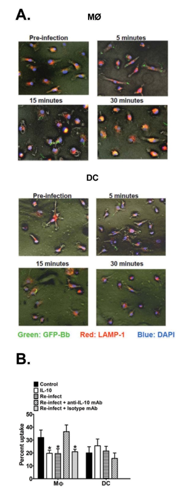
Figure 5. Effect of IL-10 on Bb uptake and trafficking by APCs.

A. MØs (upper panel) and DCs (lower panel) from B6 mice were cultured on glass coverslips and infected with GFP-expressing (green) Bb (MOI=10) at 37°C. At 5, 15, and 30 min post-infection, APCs were fixed with 4% paraformaldehyde and permeablized for staining of LAMP-1 using TRITC-labeled (red) antibodies; nuclei are stained with DAPI (blue). Immunofluorescent images (200x) are representative of triplicate samples from three separate experiments, each with at least 3 different fields of views. (B) Quantitative analysis of Bb phagocytosis by APCs. MØs and DCs from B6 mice were infected with GFP-expressing Bb as in A (above) under five different conditions: GFP-Bb only (control), recombinant IL-10 (2ng/ml) administered overnight prior to GFP-Bb infection (rIL-10), infected with non-fluorescent Bb overnight prior to GFP-Bb infection (Re-infect), same as Re-infect, but initial infection was performed in the presence of anti-IL-10 antibody (Re-infect + anti-IL-10 mAb) or an isotype control (Re-infect + isotype mAb). Percent internalization was calculated as percentage of APCs per field of view containing internalized Bb at 30 min post-Bb infection. Data represents the average of ten separate fields of views, each containing 75-150 APCs, comprising at least three separate experiments. * Indicates statistically significant values (P≤0.05) compared to cells cultured with GFP-Bb only.
