
Figure 1. Cannulae locations and muscimol spread in the prefrontal cortex and lateral entorhinal cortex.
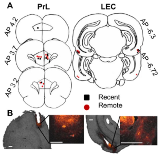
A, The rats whose cannulae tips were located in the prelimbic area of the medial prefrontal cortex (PrL; left) and lateral entorhinal cortex (LEC; right) were included in the behavioral analyses (Recent group, black; Remote group, red). The hemisphere that was implanted with a cannula was counterbalanced across rats. B, The spread of muscimol was estimated with muscimol tagged to a fluorescent molecule (red). The spread was mostly restricted to the PrL (right) or LEC (left). The inset images show magnified views of each infusion site. Scale bars indicate 500 μm.