
Figure 6. Overexpression of 14-3-3ζ downregulates synaptophysin protein level in rat hippocampal primary neurons in culture.
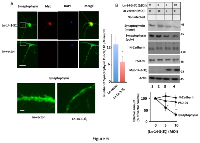
– Neurons infected with Ln-14-3-3ζ or Ln-vector were analyzed for synaptophysin protein levels by immunocytochemistry or Western blotting. (A) Immunocytochemistry. Representative immunofluorescent micrographs are of neurons infected with the indicated virus and stained for Myc (14-3-3ζ), synaptophysin, DAPI (nucleus), and merge (co-localization). The corresponding inset is shown in higher magnification in the lower panel. Quantification of synaptophysin puncta from 50 neurites in each group from three different cultures is shown on the right hand side panel. Scale bars: upper 15 μm; lower, 5 μm. *p<0.005 with respect to Ln-vector infected neurons. (B) Western blot analysis. Representative Western blots from extracts of neurons infected with Ln-14-3-3ζ or Ln-vector showing the level of indicated synaptic proteins in each culture. The relative amount of each protein was determined from the blots by normalizing the band intensity of that protein against the respective actin band. Values with standard error are an average of three determinations from three cultures. *p< 0.05 with respect to Ln-vector infected cells.