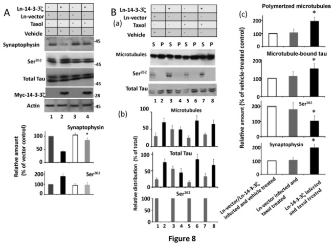
Figure 8. Microtubule stabilizing drug taxol restores synaptophysin protein levels in 14-3-3ζ overexpressing neurons.

Neurons infected with Ln-14-3-3ζ or Ln-vector were treated with the microtubule stabilizing drug taxol for 24 hr and then analyzed by Western blotting to determine the relative amounts, and then subjected to a microtubule sedimentation assay. (A) Western blot analysis. The relative amount of each protein shown in the lower panel was determined as per Figure 5. Data with standard error are the average of three determinations from three cultures. *p < 00.1 with respect to 14-3-3ζ infected and vehicle treated neurons. (B) Microtubule sedimentation assay. The microtubule sedimentation assay was performed as in Figure 4. The resulting microtubule pellet (P) and the supernatant (S) were analyzed by Western blotting and the relative distribution and relative amounts were determined as in Figure 4. (a) Western blots. (b), relative distribution. Values with S.E. are average of three determinations from three cultures. *p< 0.05 with respect to the P fraction of Ln-vector control. (c) Relative amounts. The relative amount of polymerized microtubules are relative distribution values from the microtubule pellet in panel (b) and are expressed as the % of Ln-vector control. Likewise, the relative amounts of microtubule-bound tau are the values of total tau in the microtubule pellet in panel (b) and are expressed as a % of Ln-vector control. The relative amount of Ser262 phosphorylated tau was determined by normalizing Ser262 blot by corresponding tau blot as in Figure 4. Values are an average of three determinations from three cultures. *p<0.05 with respect to Ln-vector control.
