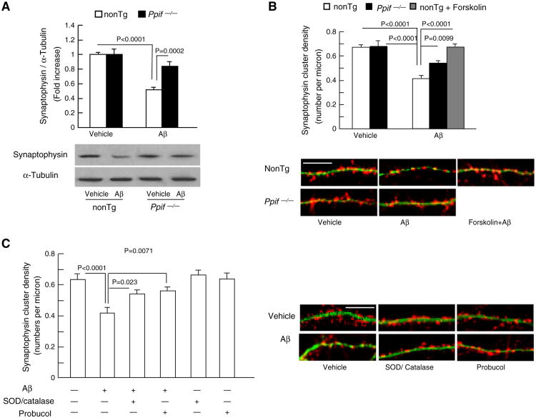
Fig. 4.
Effects of CypD deficiency and antioxidant on Aβ-induced synaptic loss. (A) Expression levels of synaptophysin in Aβ-treated nonTg and Ppif−/− neurons. Cultured neurons were treated with 5 μM oligomer Aβ1–42 for 2 h. Cell lysates were subjected to immunoblotting with synaptophysin antibody. Densitometry of the synaptophysin immunoreactive bands was normalized by α-tubulin using NIH Image J software. The lower panels are representative immunoblots for synaptophysin and α-tubulin. Data were collected from 3 to 4 independent experiments. (B) After exposure of 5 μM oligomer Aβ for 2 h, Ppif−/− neurons exhibited preserved synaptic positive clusters compared with nonTg neurons. PKA activator, forskolin (5 μM), protected nonTg neurons against Aβ-induced synaptic loss. Data were collected from 17 to 20 neurons per group. The lower panel shows representative images of synaptophysin clusters (synaptophysin, red) and dendrite (MAP2, green) staining. Scale bar = 10 μm. (C) Antioxidants attenuated Aβ-induced synaptic loss. Data are derived from 16 to 23 neurons per group in 3 independent experiments. The right panel shows representative images of synaptic staining. Synapses were visualized by synaptophysin staining (red) and dendrites were shown by MAP2 staining (green). Scale bar = 10 μm.

