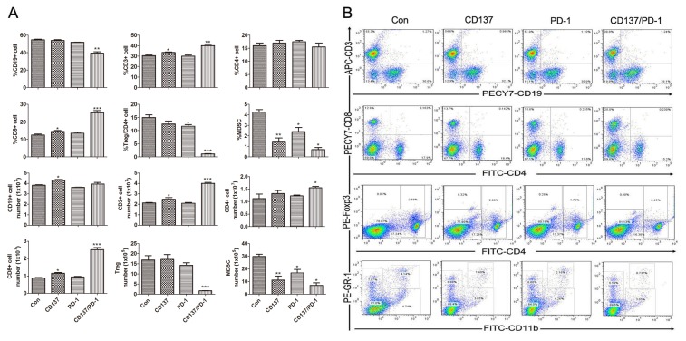
Figure 2. Analysis of lymphocyte components in spleens from mice treated with mAb combinations.
Mice (3/group) transplanted i.p. with 3 × 10⁶ ID8 cells 10 days earlier were injected i.p. with 0.5 mg of control, anti-CD137, anti-PD-1 or anti-PD-1/CD137 mAb and the mAb injection was repeated 4 days later. Seven days after the second injection, spleens were harvested and single cell suspensions prepared and stained with fluorescence labeled antibodies against markers of lymphocyte subsets prior to analysis by flow cytometry. The percentages and numbers of CD3+, CD4+, CD8+, CD19+, FoxP3+/CD4 and GR-1+CD11b+ cells in spleens are shown in (A) and representative dotplots are shown in (B). Data are presented as M ±SEM from 3 mice/group and are representative of 3 independent experiments. *P < 0.05, **P < 0.01, ***P < 0.001, The findings with anti- PD-1 or CD137 single mAbs are compared with control mAb, and the findings with anti- PD-1/CD137 mAbs are compared with both control and single mAb.

