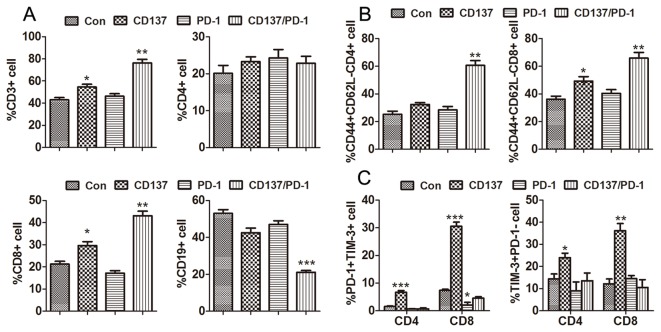
Figure 5. Analysis of peritoneal lymphocyte subsets from mice injected with the anti-PD-1/CD137 combination.
Mice (3/group) transplanted i.p. with 3 × 10⁶ ID8 cells 10 day earlier were injected i.p. twice at 4 days interval with 0.5 mg of control, anti-CD137, anti-PD-1 or anti-PD-1/CD137 mAb. Two weeks later, peritoneal lavage from treated mice was analyzed by flow cytometry for the percentage and phenotype of peritoneal lymphocytes. The percentages of CD3+, CD4+, CD8+ and CD19+ lymphocytes in peritoneal lavage and CD44+CD62L- effector/memory cells in CD4+ and CD8+ T cells are shown in (A) and (B) respectively. The percentage of PD-1+TIM-3+ and PD-1-TIM-3+ cells in peritoneal CD4+ and CD8+ T cells are shown in (C) with representative dotplots in Figure S2. Data are presented as M±SEM from 3 mice of each group and are representative of 2 independent experiments. *P < 0.05, **P < 0.01, ***P < 0.001, PD-1 or CD137 mAb compared with control mAb, PD-1/CD137 mAb compared with control and single mAb.

