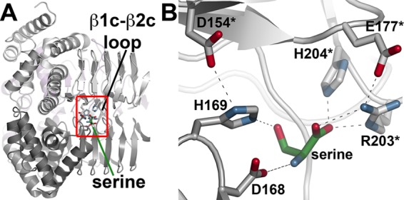
FIGURE 4.

Structure of the serine binding site. A, view of the location of serine binding between two GmSAT monomers. The flexible β1c-β2c loop and serine are indicated. The red box shows the area highlighted in B. B, the serine binding site of GmSAT. Residues contributed by each monomer are colored gray and white, respectively, with an asterisk to indicate the second monomer.
