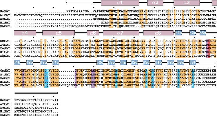
FIGURE 6.

Sequence comparison of SAT from different organisms. The amino acid sequences of the SAT from soybean (GmSAT; NP_001235640.1), A. thaliana (AtSAT; AAC37474.1), E. coli (EcSAT; NP_290190.1), H. influenzae (HiSAT; NP_438764.1), and E. histoyltica (EhSAT; BAA82868.1) are shown. The secondary structure of GmSAT is shown above the alignment with α-helices (rose) and β-strands (blue) indicated. White boxes indicate disordered regions of the GmSAT structure. Highly conserved residues (orange), residues in the serine binding site (purple), residues in the CoA binding site (blue), and the catalytic histidine (red) in the SAT are highlighted.
