FIGURE 3.

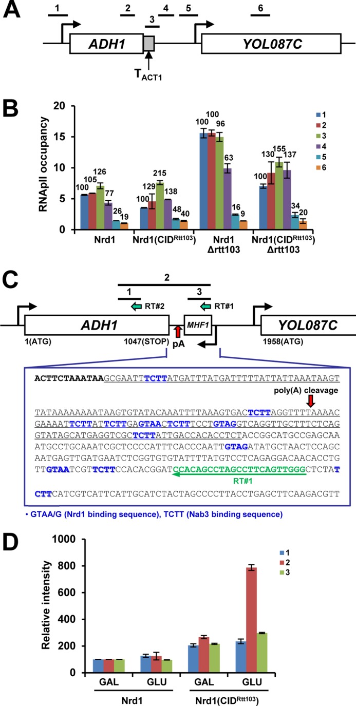
Nrd1(CIDRtt103) alters termination at ADH1. A, schematic diagram of ADH1 with ACT1 terminator. ADH1 3′-flanking region (183 bp as underlined in C, TADH1) was replaced with ACT1 terminator (TACT1, 273 bp). Positions of PCR primers are shown above the diagram. B, termination of ADH1-TACT1 becomes defective in Nrd1(CIDRtt103) strains, indicating that TACT1 may contain cis-acting element(s), responding to Nrd1(CIDRtt103). C, ORF map and sequences downstream of ADH1 are shown. ADH1 has multiple Nrd1/Nab3-binding sites near the pA site: ADH1 ORF in bold, Nrd1/Nab3-binding sites in blue, RT primer (RT#1) in green, and TADH1 (replaced with TACT1 as in A) is underlined. Reverse transcription using RT#1 primer was performed with RNAs from WT (Nrd1) and Nrd1(CIDRtt103) cells grown in either galactose or glucose (24 h) media to deplete Rrp41. Similar analysis was done using RT#2 primer for loading control and band normalization. Bars above the diagram denote PCR products subsequently generated. D, quantification of RT-PCR results. The band intensities of PCR products from the WT-galactose sample were set to 100% and those from the rest of samples were calculated in relative amounts. Results are shown from three repetitions. Error bars represent S.E.
