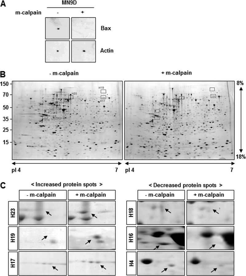
FIGURE 2.
Representative 2DE gel images of putative calpain substrates. A, mini-2DE analysis was carried out to confirm that calpain effectively degraded Bax, one of its known substrates (15, 16). Lysates (100 μg) obtained from MN9D cells were loaded on 7-cm Drystrips and separated by IEF ranging from pI 4 to 7. Next, samples in the ministrips were incubated with 5.55 units of m-calpain for 2 h at 37 °C, subjected to 12% SDS-PAGE, and probed with anti-Bax antibody. Actin was used as a loading and a negative control. B, representative gel images are shown for samples treated with or without m-calpain and further processed for 2DE (24 cm, pI 4–7, and 8–18%). Briefly, total protein lysates (1.5 mg) obtained from MN9D cells were separated by IEF and incubated with or without 83.25 units of m-calpain for 2 h at 37 °C. After separation by SDS-PAGE, the gels were stained with 0.1% Coomassie Brilliant Blue G-250. Approximately 1,500 protein spots ranging from 10 to 100 kDa were routinely detected. C, comparative close-up views of the typically altered protein spots boxed in Fig. 2B, before and after calpain treatment. Arrows indicate the protein spots that were either increased (left) or decreased (right) following calpain treatment. Detailed molecular information for these protein spots is shown in Table 2.

