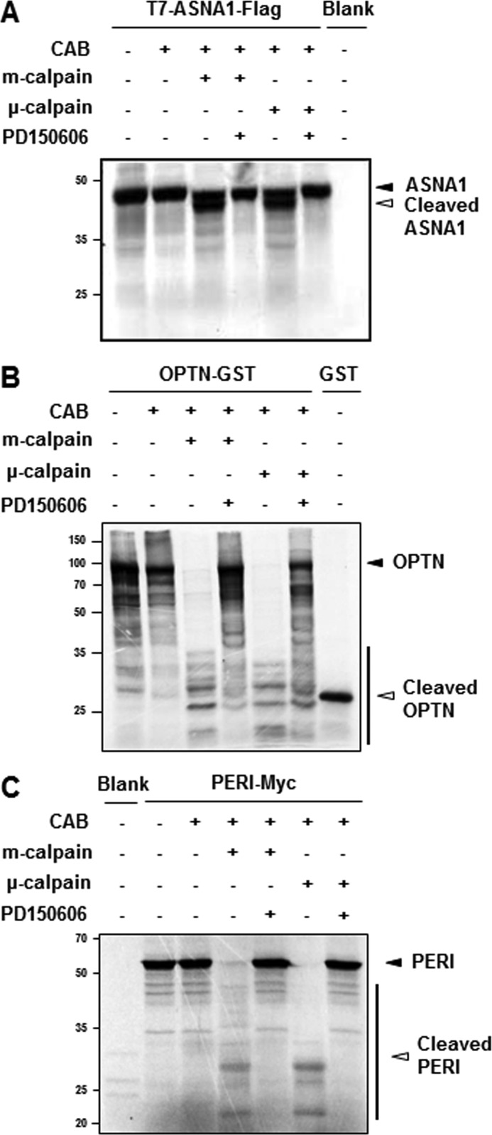
FIGURE 3.
In vitro calpain cleavage assays. Among the altered protein spots identified as putative calpain substrates, T7-ASNA1-FLAG (A), OPTN-GST (B), and PERI-Myc (C) were radiolabeled with [35S]methionine using an in vitro transcription/translation kit. Reactants were incubated for 2 h at 37 °C with the indicated amount of recombinant calpains (5.55 and 3.95 units for m- and μ-calpain, respectively) in the presence or absence of a calpain inhibitor, PD150606 (500 μm) in a calpain activation buffer (CAB). After incubation, all samples were separated by SDS-PAGE and subjected to autoradiography. Closed arrowheads indicate full-length proteins, and open arrowheads represent cleaved fragments. Autoradiographic images are representative from at least three independent experiments.

