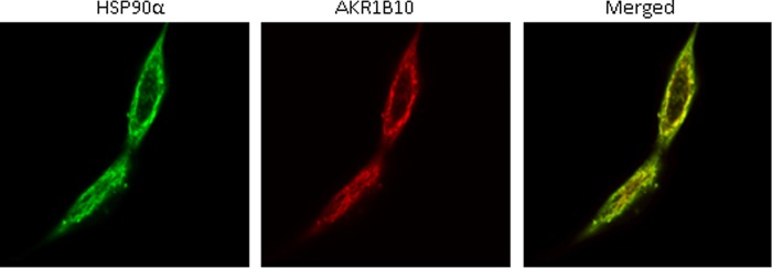
FIGURE 2.

Fluorescent colocalization. Fluorescent immunocytochemistry and laser confocal imaging were carried out as described under “Materials and Methods.” The secondary antibodies against the primary antibodies to HSP90α and AKR1B10 were labeled with green FITC and red Rhodamine, respectively, and the merged yellow image indicates the colocalization of AKR1B10 and HSP90α in cytosol.
