
FIGURE 5.
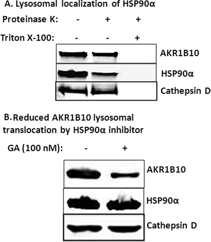
Lysosomal localization of HSP90α and AKR1B10. A, proteinase K protection. HCT-8 cells (5 × 10⁷) were broken down using a Dounce homogenizer, and lysosomes were isolated for proteinase K protection assays as described under “Materials and Methods.” Triton X-100 (0.5%) was added in a group to destroy biomembranes. B, HSP90α inhibition. HCT-8 cells (2.5 × 10⁷) were incubated for 12 h with 100 nm HSP90α inhibitor GA. Lysosomes were isolated, and AKR1B10 and HSP90α in the lysosomes were detected by Western blot analysis.