Figure 3.

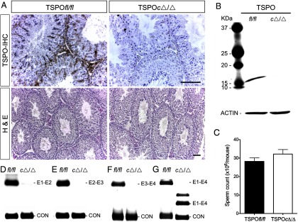
TSPO deletion in Leydig and Sertoli cells does not affect spermatogenesis. A, Immunohistochemical (IHC) localization showing complete absence of TSPO in Leydig and Sertoli cells of TSPOcΔ/Δ testes. Hematoxylin and eosin (H&E) staining showing unaltered seminiferous tubule morphology and spermatogenesis in TSPOcΔ/Δ testes (n = 5). Scale bars, 50 μm. B, Western blot showing absence of TSPO in TSPOcΔ/Δ testis tissue (n = 5); β-actin is shown as the loading control. C, Cauda epididymal sperm counts were not significantly different between TSPOfl/fl and TSPOcΔ/Δ mice (mean ± SEM; n = 5/group). D–F, Testis cDNA from TSPOfl/fl and TSPOcΔ/Δ mice examined for amplification products from exons 1 and 2 [250 bp] (D); exons 2 and 3 [241 bp] (E); exons 3 and 4 [424 bp] (F); exons 1–4 [711 bp in TSPOfl/fl and 361 bp in TSPOcΔ/Δ]. For all RT-PCR, glyceraldehydes-3-phosphate dehydrogenase was used as a control (CON).
