Figure 7.
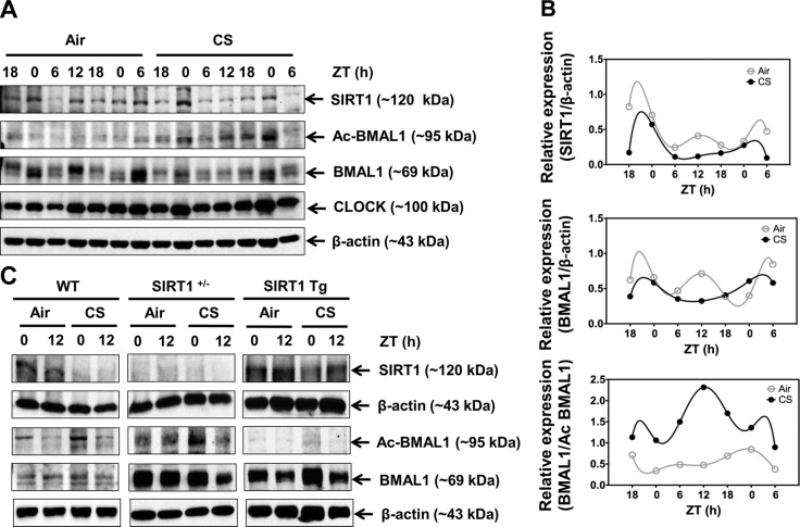
Circadian rhythm of SIRT1 expression in lung tissue was disrupted by CS exposure, resulting in elevated BMAL1 acetylation. A) Immunoblot analysis of SIRT1, total BMAL1, acetylated BMAL1 (Ac BMAL1), and CLOCK in lung tissue homogenates from mice after 10 d of air or CS exposure. Images are representative of ≥2 separate experiments. B) Oscillation patterns of SIRT1, BMAL1, and Ac BMAL1 protein as shown by immunoblot analysis. After densitometric analysis, levels of SIRT1 and BMAL1 were both normalized against β-actin, and Ac BMAL1 was normalized against total BMAL1. Data from air- and CS-exposed samples are representative of the immunoblot data, and were analyzed with nonlinear regression, as described in Materials and Methods (R2=1 for all data). C) Genetic manipulation of SIRT1 regulated BMAL1 acetylation in response to CS in the lungs. Immunoblot analysis of SIRT1, BMAL1, and acetylated BMAL1 performed in lung tissue homogenates from SIRT1 heterozygous knockout (SIRT1+/−) and SIRT1-overexpressing (SIRT1 Tg) mice following 3 d of air or CS exposure. Images are representative of ≥2 separate experiments. Reassembly of noncontiguous gel lanes is demarcated by white spaces and boxes, aligned so as to reflect the overall representative data. Data from air- and CS-exposed tissue are shown as means ± se (n=2–3/group) for each time point.

