Figure 1.

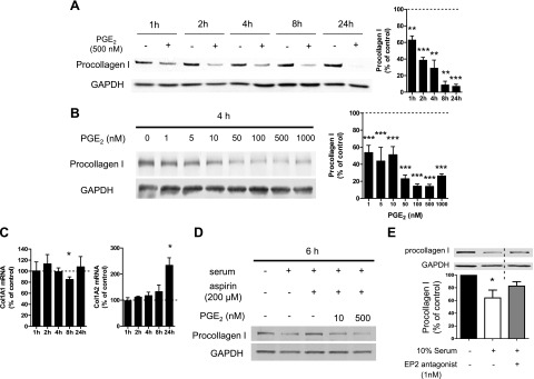
Rapid suppression by PGE2 of procollagen I protein expression in normal adult lung fibroblasts. A–C) After overnight serum starvation, cell culture medium was changed to fresh serum-free medium, and normal adult lung fibroblasts were then incubated with or without PGE2 (at 500 nM in A and at various concentrations in B). A, B) Time course (A) and dose dependency (B) of the effect of PGE2 on procollagen I protein expression. At each time point after addition of PGE2 at the indicated concentrations, cell lysates were harvested, and procollagen I levels in the lysates were determined by immunoblot analysis with densitometry. Left panel: representative immunoblot. Right panel: results of densitometric analysis of procollagen I levels from 3–5 experiments (A) or 3 experiments (B). After normalization to GAPDH, the procollagen I level in PGE2-treated cells was expressed relative to that in cells without PGE2 (expressed as 100%, dashed line) at each time point in each experiment. Data represent means ± sem. C) PGE2 did not suppress collagen I mRNA expression. At the indicated time points, ColIα1 (left panel) and ColIα2 (right) mRNA levels in cell lysates were determined by semiquantitative real-time RT-PCR and then were expressed relative to the level in the no-PGE2 control condition (expressed as 100%, dashed line) at each time point in each experiment. Data are expressed as means ± sem from 4–5 (left panel) or 3–4 (right panel) experiments. *P < 0.05, **P < 0.01, ***P < 0.001 vs. control fibroblasts without PGE2 treatment. D, E) Serum decreased procollagen I protein expression via the PGE2-EP2 pathway. After cells were serum starved overnight, serum-free medium was removed, and cells were incubated in serum-containing medium, with or without aspirin (200 μM) (or its vehicle DMSO at a final concentration of 0.2%) in the absence or simultaneous presence of PGE2 (10 or 500 nM; D), or with or without the EP2 antagonist PF-04418948 (1 nM; E) for 6 h. Then collagen I protein levels in the cell lysates were determined by immunoblot analysis. Immunoblot representative of 3 experiments is shown. Lanes separated by the vertical dashed line were from the same blot, but were not contiguous in the original gel. After normalization to GAPDH, the procollagen I level in PGE2-treated cells was expressed relative to that in cells treated without serum (expressed as 100%) in each experiment (E). Data represent means ± se. *P < 0.05 vs. control fibroblasts without serum treatment.
