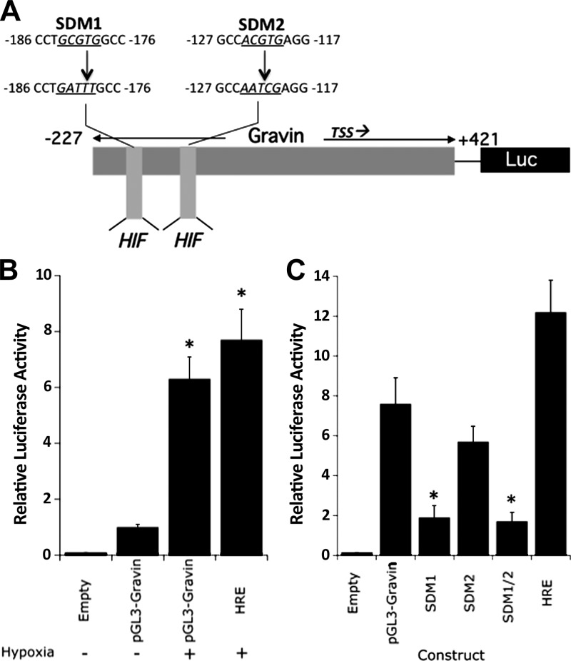
Figure 3.
Cloning and functional analysis of the gravin promoter. A) Map of cloned gravin promoter and luciferase constructs utilized here. Relative positions of each clone are annotated, as well as the transcription start site (TSS). Putative HIF binding sites (HIF) are shown, as are the sequences of the site-directed mutants SDM1 and SDM2. B) HMEC-1 monolayers were transfected with plasmids expressing sequence corresponding to full length gravin (pGL3-gravin) or the positive control plasmid encoding the HRE from the erythropoietin gene (HRE). Transfected cells were exposed to hypoxia or normoxia for 24 h and assessed for luciferase activity. Data are means ± sem, n = 3. *P < 0.01 vs. normoxia. C) HMEC-1 monolayers were transfected with plasmids expressing sequence corresponding to full length gravin (pGL3-gravin), SDM1, SDM2, or SDM1/2, or the positive control plasmid encoding the HRE from the erythropoietin gene (HRE). Transfected cells were exposed to hypoxia or normoxia for 24 h and assessed for luciferase activity. Data are means ± sem, n = 3. *P < 0.01 vs. normoxia.

