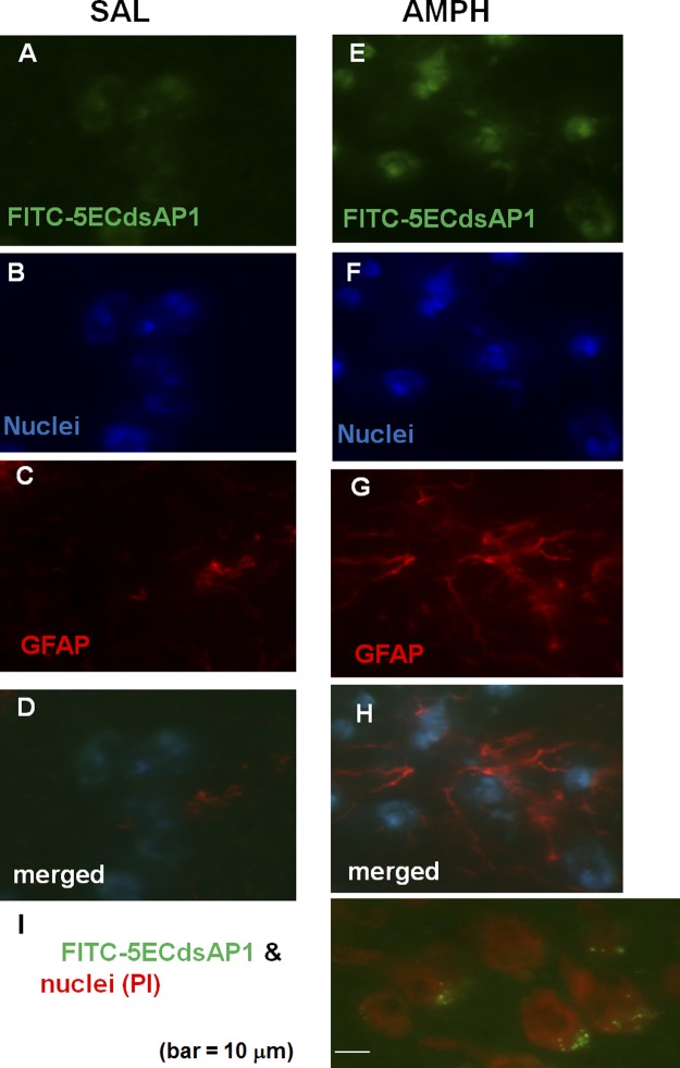
Figure 2.
Uptake and distribution of aptamers. To examine for cell-specific uptake of AP-1 aptamer, we delivered FITC-5ECdsAP1 (120 pmol/kg, i.c.v. injection). We then administered saline (SAL; A–D) or amphetamine (AMPH; 4 mg/kg, i.p. injection; E–I) 3 h later (n=2 each); frozen brain samples were obtained 4 h after saline or amphetamine (15). A, E) Brain tissue was stained for FITC-5ECdsAP1 detection. B, F) Brain tissue was stained for nuclei detection. C, G) Brain tissue was treated with fresh 4% paraformaldehyde to label GFAP expressed by astroglia [Cy3-labeled antibodies against GFAP (ab7260; Abcam), and Hoechst (purple) stain for nucleic acids]. D, H) Merged images. I) FITC-5ECdsAP1 was located near the neuronal nuclei (∼10 μm in diameter) stained with propidium iodide. Scale bar = 10 μm.

