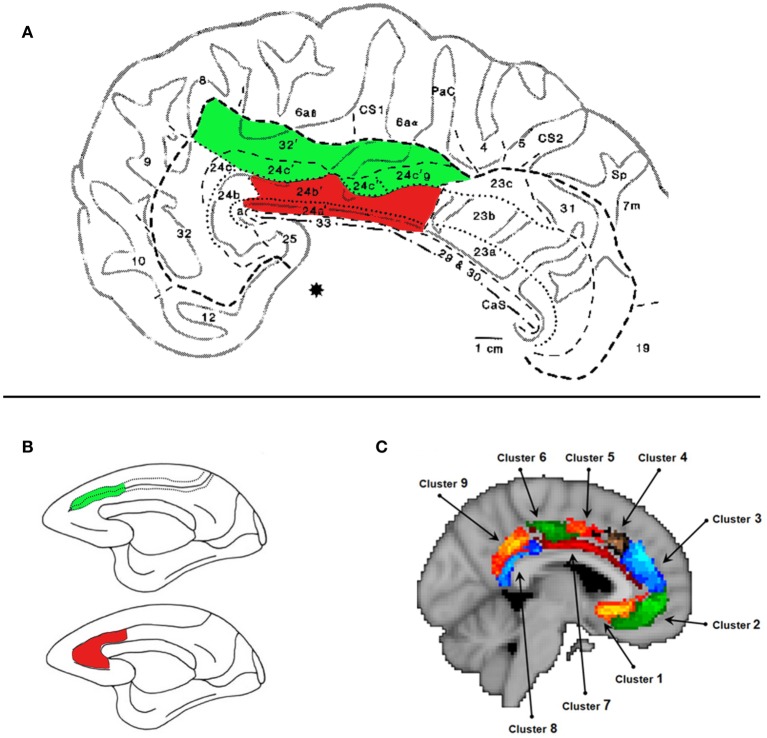
Figure 1.
The Midcingulate Cortex (MCC). (A) Cytoarchitecture of the MCC taken from Vogt et al. (1995). The areas shaded in green lie in the MCCs. The areas shaded in red lie on the MCCg. We argue that this area is engaged when processing information about others' decisions. Specifically we argue that areas 24a' and 24b', which lie on gyral surface of the cingulate cortex, extending on average 22 mm posterior to and 30 mm anterior to the anterior commisure denoted by (*). (B) Lesion site of the MCCg and ACCg (red) and the MCCs and the ACCs (green) from Rudebeck et al. (2006). The lesions that affected the gyrus caused disruptions to social behavior and disrupted the processing of social stimuli. (C) Subdivisions of the MCC and ACC according resting-state connectivity (Beckmann et al., 2009). Cluster 7 shown in dark red corresponds, broadly, to the MCCg.

