Figure 2.
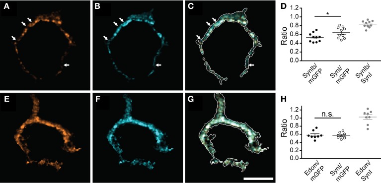
Multiple synapsin isoforms are co-expressed within the same presynaptic domains. Calyces stained with isoform-specific antibodies (A,E), and pan-synapsin I antibody (B,F). Overlap with calyx outline, based on the mGFP-expression delineates the membrane of the calyx in the merged images (white outline, C,G). The volume of the entire calyx was calculated from mGFP labeling (see Materials and Methods). (A–C) synapsin Ib. Arrows indicate pan-synapsin I positive clusters with no detectable synapsin Ib. (D) Ratios of the presynaptic volume occupied by synapsin Ib and synapsin I. Synapsin Ib occupied 0.53 ± 0.03, and synapsin I occupied 0.64 ± 0.04 of the presynaptic volume (N = 10 presynaptic terminals, Student's t-test, P = 0.04). Overlap of synapsin Ib and synapsin I signal was 0.83 ± 0.02. (E–G) Synapsin E-domain revealing all isoforms containing E-domain (i.e., synapsin Ia, IIa, IIIa). (H) Ratios of the presynaptic volume occupied by synapsin E domain and synapsin I. Synapsin E domain occupied 0.58 ± 0.04, synapsin I occupied 0.57 ± 0.03 of the presynaptic volume (N = 8 presynaptic terminals, Student's t-test, P = 0.81). The overlap of synapsin E domain and synapsin I volume was 1.03 ± 0.04. Images represent single confocal planes in pseudocolors. Scale bar is 10 μm. *P < 0.05.

