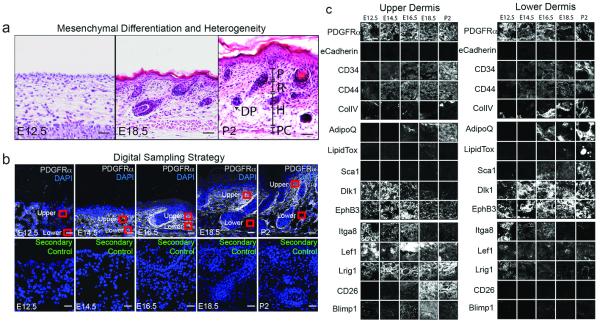
Figure 1. Morphological and molecular markers of embryonic and postnatal fibroblasts.

(a-c) Sections of mouse back skin. (a) H&E staining. P: papillary dermis; R: reticular dermis; H: hypodermis; P: panniculus carnosus; DP: dermal papilla. (b) 10μm sections immunostained with PDGFRa antibodies and secondary antibody control with DAPI nuclear counterstain. Red squares show areas digitally sampled for the tissue screen. (c) Tissue screen of upper and lower dermis. 3 biological replicates for each stain were performed. Images are representative samples of sections shown Fig. E1, E2. Scale bars: 50 μm.
