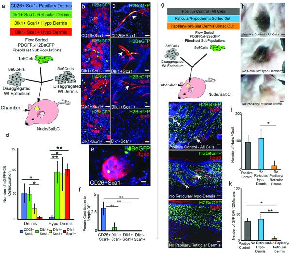
Figure 2. Skin reconstitution assays.
(a) Experimental set up for (b-f). (b-c) Grafts immunostained with DAPI counterstain (blue). SG: sebaceous gland. Arrows: APM. (d-f) Contribution of PDGFRaH2BeGFP cells to dermal compartments. (g) Experimental set up for (h-k). (h) Macroscopic views of grafts. (i) Contribution of GFP+ cells to grafts. Arrows: GFP+ DP. (j) Hair follicles per graft. (k) GFP+ DP per 120 microns graft. N=3 biological replicates per experiment. *P <0.05; **P <0.005. Horizontal whole mounts shown. Scale bars: (b-c) 40 μm (e) 30 μm (h) 2.5mm (i) 50 μm.

