
Figure 4.
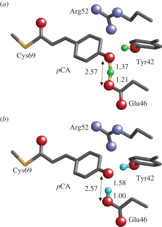
Geometry of the photoactive site in PYP. Only the H atom position of the H-bonds between Tyr42 and pCA, and between Glu46 and pCA are shown (green or cyan spheres). O and N atoms of the side chains are depicted as red and blue balls, respectively. (a) Neutron diffraction analysis [39] (PDB; 2ZOI). (b) QM/MM optimized structure [45] based on the X-ray crystal structure (PDB; 2ZOH). (Online version in colour.)