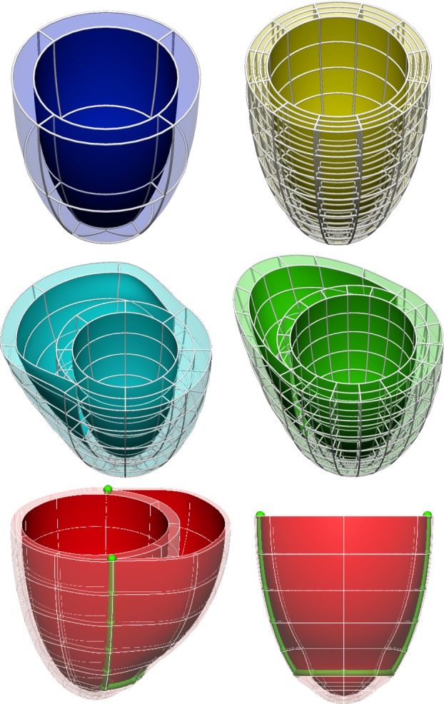
Figure 2.

Examples of template meshes of the LV and the BiV anatomy with different resolutions and dimensions of the right ventricular blood pool. Bottom row highlights in green the squared insertion line of the RV into the LV (line that joins the ventricular cusps referred in [19]).
