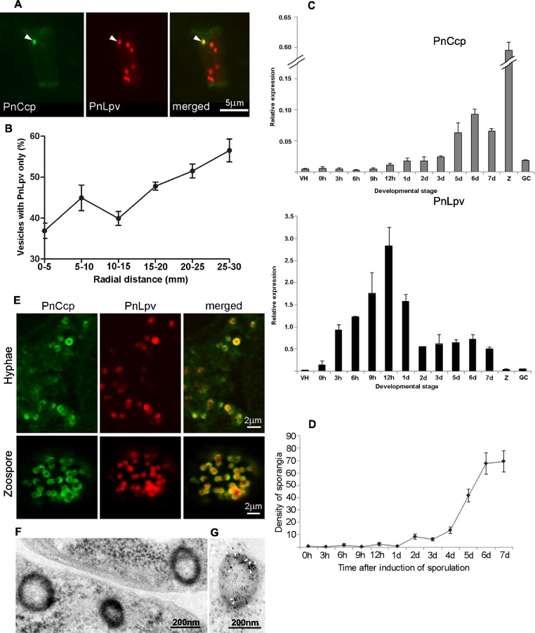
Figure 3. PnCcp and PnLpv synthesis and packaging into large peripheral vesicles.
(A) Double-immunofluorescence labelling with PnCcpCpep and Lpv-1 antibodies shows only one (arrowhead) of seven vesicles in a hyphal fragment from the leading edge of mycelium growing on nutrient agar contains PnCcp in addition to PnLpv proteins. (B) Quantitation of vesicles that contain PnLpv only in hyphae sampled at 5-mm intervals across a mycelial colony from its centre (0–5 mm) to the advancing edge (25–30 mm). Bars indicate s.e.m. (n = 3). (C) qPCR quantitation of PnCcp and PnLpv transcript levels in vegetative hyphae (VH), sporulating hyphae (0 h to 7 days), zoospores (Z) and 3-h germinated cysts (GH). Expression levels are relative to the normalising gene, WS041. Bars indicate s.e.m. (n = 3). (D) Density of sporangia (per microscope field of view) in P. nicotianae mycelia growing in liquid culture after induction of sporulation. Bars show s.e.m. (n = 3). (E) Double-immunofluorescence labelling with PnCcpCpep and Lpv-1 antibodies of P. nicotianae sporulating hyphae and zoospores. Confocal microscope optical sections show that PnCcp (green) is often restricted to an outer zone within the large peripheral vesicles while PnLpv (red) occurs throughout the vesicles. (F, G) Large peripheral vesicles in cleaving sporangia of P. nicotianae prepared by plunge-freezing and freeze-substitution. Ultrastructural analysis reveals an outer shell of electron-dense material (F) which is labelled by PnCcpCpep-goat anti-rabbit-10 nm gold (arrowheads). Lpv-1-goat anti-mouse-5 nm gold labels throughout the vesicle (G).

