
Figure 10. Putative IPSC colony showing mRFP1-positive and mRPF1-negative cells.
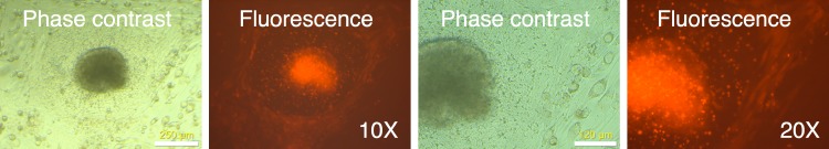
mRFP-negative colonies were picked using a pulled glass Pasteur pipet as described in Materials and Methods and placed in a well with mouse feeder cells. The following day, the well was observed under phase contrast and fluorescence microscopy.