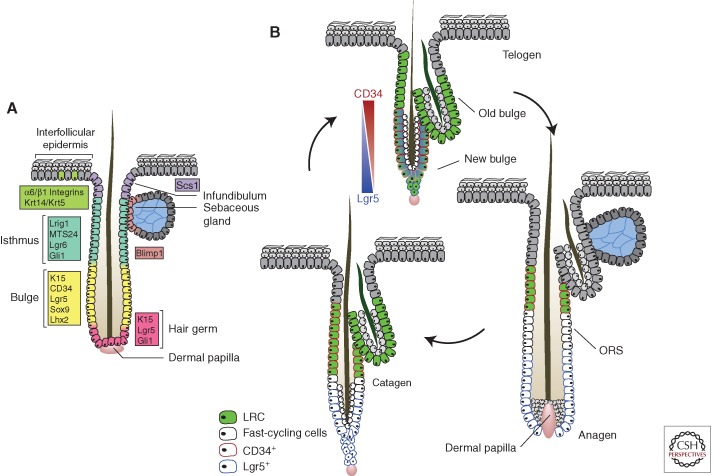
Figure 3.
Hair follicle stem cell dynamics. (A) Summary of the expression of proposed stem cell markers in the hair follicle. The extent to which these markers identify functionally distinct populations has yet to be fully resolved. (B) Stem cell proliferation and migration in the hair cycle. During anagen, cells from the lower bulge region start proliferating, contributing to the formation of the outer root sheath (ORS). Genetic label retaining shows that some bulge cells remain quiescent (green), whereas others migrate into the lower follicle, losing their stem cell properties, and proliferate (white). In catagen, the proliferating cells return to the bulge where they provide a niche sustaining the quiescent stem cells through telogen and into the next hair cycle. Marker expression changes dynamically through the cycle. CD34 is expressed in the telogen bulge and retained by quiescent stem cells throughout the cycle. Lgr5 expression overlaps with CD34 in telogen, but is localized to the proliferating cells in the lower follicle in anagen. The functional significance of the changes in Lgr5 expression is unclear. (Figure based on data from Jaks et al. 2008 and Hsu and Fuchs 2012.)

