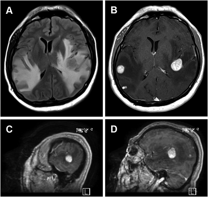
Figure 1.
Brain MRI of patient with multiple intracerebral metastases from lung adenocarcinoma. A, Axial T2-FLAIR MRI shows extensive abnormal hyperintensity within the bilateral temporoparietal subcortical white matter and left internal and external capsules, with effacement of overlying cortical sulci and underlying lateral ventricles as well as rightward shift of midline structures. B, Axial T1-postgadolinium MRI shows multiple homogenously avidly enhancing round metastases. Sagittal T1-postgadolinium MRI shows the proximity of the large right-sided (C) and left-sided (D) metastases to the superior temporal gyri. FLAIR indicates fluid attenuated inversion recovery; MRI, magnetic resonance imaging.

