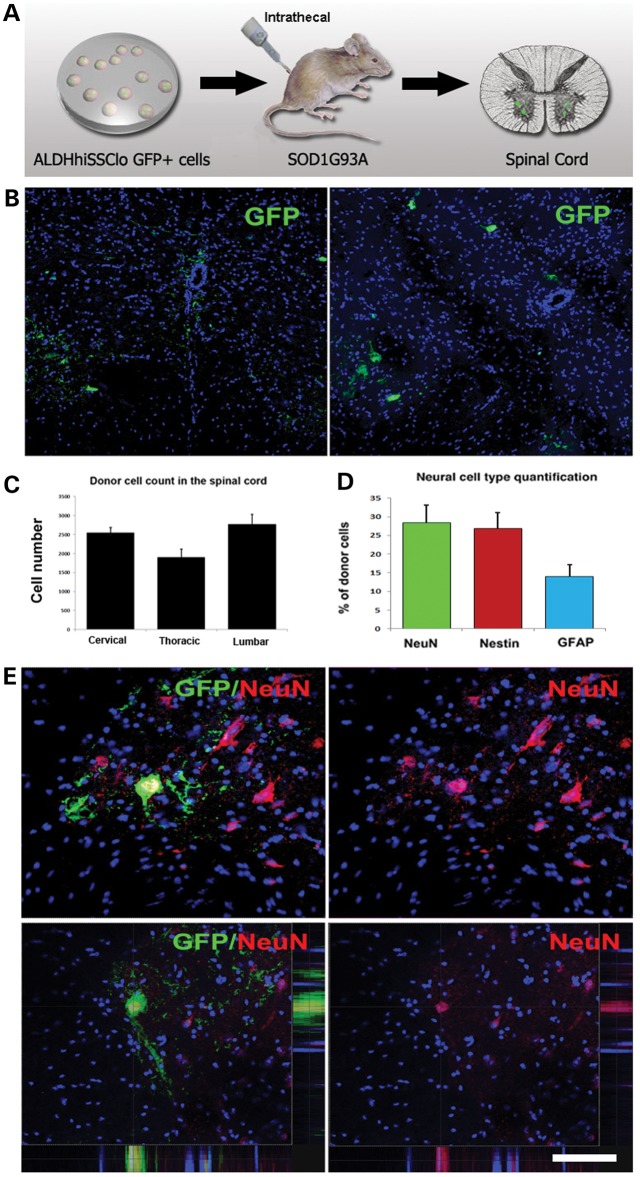
Figure 2.
iPSC-derived NSCs migrate and engraft into the spinal cords of SOD1G93A mice after intrathecal and systemic transplantation. (A) Experimental design: GFP-NSCs (1 × 10⁶ cells) were delivered by intrathecal injection into SOD1G93A mice at 90 days of age, with two additional injections at 105 and 120 days. (B) Donor GFP+ cells were detected in the spinal cord, particularly in the anterior horns. (C) Quantification of GFP-donor cells in the cervical, thoracic and lumbar spinal cord. Error bars indicate the SD. (D) Quantification of the phenotype acquired by the donor cells revealed the presence of cells with an undifferentiated phenotype (nestin), neuronal (NeuN) phenotype and glial cells (GFAP). Error bars indicate the SD. (E) Representative images of cells acquiring a neuronal phenotype, as indicated by positivity for NeuN (red) and GFP (green). Nuclei are counterstained with DAPI (blue signal). Scale bars: (B) 150 µm and (E) 50 µm.

