Figure 5.
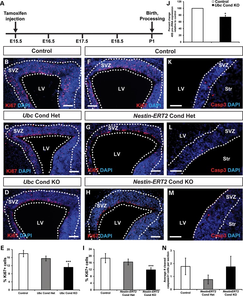
P1 Chd7 conditional knockout mice display reduced SVZ proliferation. (A) Schematic showing the tamoxifen dosing regimen where pregnant females received a single intraperitoneal injection of tamoxifen (0.2 mg/gbw) at E15.5. (B–D), (F–H) and (K–M) Representative coronal sections showing immunofluorescence for the proliferative marker Ki67 or apoptosis marker Casp3 with DAPI counterstain in the SVZ of P1 control (B, F and J), Ubc Cond Het (C), Ubc Cond KO (D), Nestin-ERT2 Cond Het (G and K) and Nestin-ERT2 Cond KO (H and L) mice. (E and I) Quantification showing the percentage of Ki67+ cells relative to the total number of DAPI-stained cells in control, Ubc Cond Het and Ubc Cond KO (E), and Nestin-ERT2 Cond Het and Nestin-ERT2 Cond KO (I) P1 SVZ. The percentage of Ki67+ cells are reduced by 37 and 38% in the SVZ of Ubc Cond KO and Nestin-ERT2 Cond KO P1 mice, respectively, compared with controls. (J) Quantitative PCR shows that 45S pre-rRNA is decreased by 26% in Ubc Cond KO SVZ compared with controls. (K–N) There is no change in the number of Casp3+ cells per section in Nestin-ERT2 Cond Het (L) and Nestin-ERT2 Cond KO (M) P1 SVZ compared with control (K). Scale bars in (B–D), (F–H): 75 μm, (K–M): 50 μm. Error bars in (E, I, J and N) indicate SEM (n = 3 per genotype). *P < 0.05, ***P < 0.001 by unpaired Student's t-test.

