Figure 7.
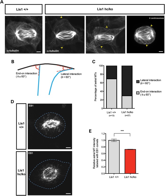
Loss of LIS1 causes aberrant interactions between astral MT plus-ends and the cell cortex. (A) The astral MTs in early anaphase from Lis1hc/ko MEFs and WT MEFs stained with α-tubulin after glutaraldehyde fixation. Arrow: aberrant astral MT tips in Lis1hc/ko MEFs. (Right panel) Misattachment of astral MTs to the opposite polar cortex in Lis1hc/ko MEFs harboring extra centrosomes (four centrosomes). (B) Schematic representation of interaction between astral MTs and the cell cortex–end-on interaction (θ° ≥ 60) versus lateral interaction (θ° < 60). (C) Quantification of types of astral MT interaction with the cell cortex in MEFs. (D) Localization of MT plus-ends stained with EB1 (MT plus-end binding protein) in metaphase of Lis1hc/ko MEFs and WT MEFs. Blue dashed lines indicate the cell membranes. Scale bars: 5 μm. (E) Relative intensity of astral MTs labeled with EB1 in metaphase-arrested MEFs (more than eight cells were analyzed from each genotype). Asterisk in (E): ***P < 0.001 by Student's t-test.

