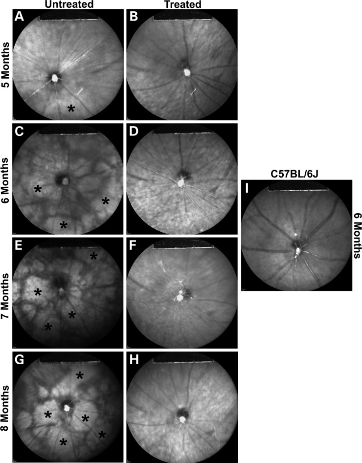
Figure 4.
Delay of retinal pigment epithelial atrophy after AAV2/8(Y733F)-Rho-Pde6α transduction. Representative infrared (IR) images of an untreated Pde6αD670G mutant eye at 5 (A), 6 (C), 7 (E) and 8 (G) months of age compared with the fellow-treated Pde6αD670G mutant eye at 5 (B), 6 (D), 7 (F) and 8 (H) months of age. Images for all time-points are taken from the same mouse. Representative IR image of a C57BL/6J control mouse at 6 months of age (I). Increased IR reflectance represents RPE atrophy (*). IR imaging was obtained at 790 nm absorption and 830 nm emission using a 55° lens. Images were taken of the central retina, with the optic nerve located at the center of the image and the site of the subretinal bleb along the lower left-hand quadrant.

