. 2013 Dec 24;2:e01503. doi: 10.7554/eLife.01503

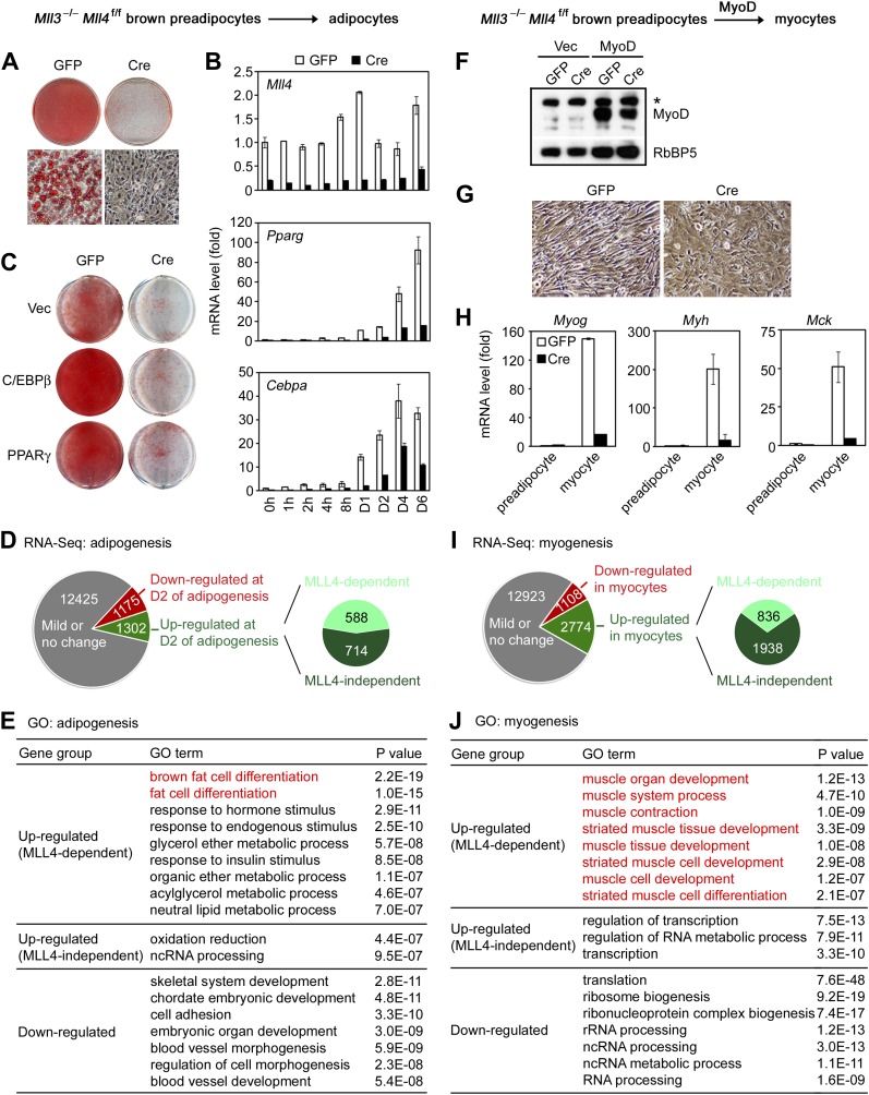
Figure 2. MLL4 controls induction of cell-type-specific genes during differentiation.

(AE) Adipogenesis of Mll3−/−Mll4f/f brown preadipocytes. (A and B) MLL4 is required for adipogenesis. Immortalized Mll3−/−Mll4f/f brown preadipocytes were infected with adenoviral GFP or Cre, followed by adipogenesis assay. (A) 6 days after induction of differentiation, cells were stained with Oil Red O. Upper panels, stained dishes; lower panels, representative fields under microscope. (B) qRT-PCR of Mll4, Pparg and Cebpa expression at indicated time points of adipogenesis. Quantitative PCR data in all figures are presented as means ± SD. D1, day 1. (C) MLL4 is required for C/EBPβ- and PPARγ-stimulated adipogenesis. Mll3−/−Mll4f/f brown preadipocytes were infected with retroviruses expressing vector (vec), C/EBPβ or PPARγ. After hygromycin selection, cells were infected with adenoviral GFP or Cre, followed by adipogenesis assay. (DE) MLL4 is required for induction of cell-type-specific genes during adipogenesis. Adipogenesis was done as in (A). Cells were collected before (day 0) and during (day 2) adipogenesis for RNA-Seq. (D) Schematic of identification of MLL4-dependent and -independent up-regulated genes during adipogenesis. The threshold for up- or down-regulation is 2.5-fold. (E) Gene ontology (GO) analysis of gene groups defined in (D). (FJ) MLL4 is required for MyoD-stimulated myogenesis. Immortalized Mll3−/−Mll4f/f brown preadipocytes were infected with retroviruses expressing Vec or MyoD. After hygromycin selection, cells were infected with adenoviral GFP or Cre, followed by myogenesis assay. (F) Western blot analysis of MyoD expression before differentiation. RbBP5 was used as a loading control. The asterisk indicates a non-specific band. (G) 5 days after induction of differentiation, cell morphologies were observed under microscope. (H) qRT-PCR analysis of myogenic gene expression after differentiation. (I and J) MLL4 is required for induction of cell-type-specific genes during myogenesis. Brown preadipocytes and myocytes were collected for RNA-Seq. (I) Schematic of identification of MLL4-dependent and -independent up-regulated genes during myogenesis. The threshold for up- or down-regulation is 2.5-fold. (J) GO analysis of gene groups defined in (I).

DOI: http://dx.doi.org/10.7554/eLife.01503.006

Figure 2.

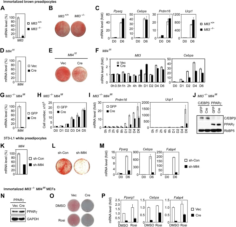
Figure 2—figure supplement 1. MLL4 is required for adipogenesis.

Figure 2—figure supplement 1.

(AC) MLL3 is dispensable for adipogenesis of brown preadipocytes. Brown preadipocytes isolated from Mll3+/+ and Mll3−/− E18.5 embryos were immortalized by retroviruses expressing SV40T. Cells were induced for adipogenesis for 6–7 days. (A) qRT-PCR analysis of Mll3 expression before adipogenesis. (B) Oil Red O staining after adipogenesis. (C) qRT-PCR analysis of gene expression before (D0) and after (D6) adipogenesis. (DF) Single KO of Mll4 leads to reduced adipogenesis. (D) qRT-PCR analysis of Cre-mediated Mll4 deletion before adipogenesis. (E) Oil Red O staining after adipogenesis. (F) qRT-PCR analysis of gene expression at indicated time points during adipogenesis. (G and H) Deletion of Mll4 does not affect growth of immortalized preadipocytes. (G) qRT-PCR analysis of Mll4 deletion in Cre-infected Mll3−/−Mll4f/f preadipocytes. (H) Cell growth curves. (I) qRT-PCR of BAT-specific Prdm16 and Ucp1 expression during adipogenesis. (J) Western blot analyses of retroviral C/EBPβ and PPARγ expression in adenoviral GFP- or Cre-infected Mll3−/−Mll4f/f brown preadipocytes before adipogenesis. RbBP5 was used as a loading control. (KM) Knockdown of Mll4 inhibits adipogenesis of 3T3-L1 white preadipocytes. 3T3-L1 cells were infected with lentivirus shRNA targeting Mll4 or control (Con) virus, followed by adipogenesis assay. (K) qRT-PCR confirmation of Mll4 knockdown before adipogenesis. (L) Oil red O staining after adipogenesis. (M) qRT-PCR analysis of gene expression before (D0) and after (D6) adipogenesis. (NP) MLL4 is required for PPARγ-stimulated adipogenesis of MEFs. 3T3-immortalized Mll3−/−Mll4f/f MEFs were infected with retroviruses MSCVhyg-PPARγ, followed by infection of MSCVpuro-Cre. Adipogenesis was induced with PPARγ ligand Rosiglitasone (Rosi) or vehicle DMSO alone. (N) Western blot analysis of retroviral PPARγ expression before adipogenesis. GAPDH was used as a loading control. (O) Oil red O staining and (P) qRT-PCR analysis of gene expression after adipogenesis.
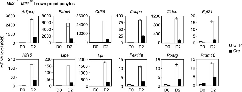
Figure 2—figure supplement 2. Confirmation of RNA-Seq data by qRT-PCR.

Figure 2—figure supplement 2.