. 2013 Dec 24;2:e01119. doi: 10.7554/eLife.01119

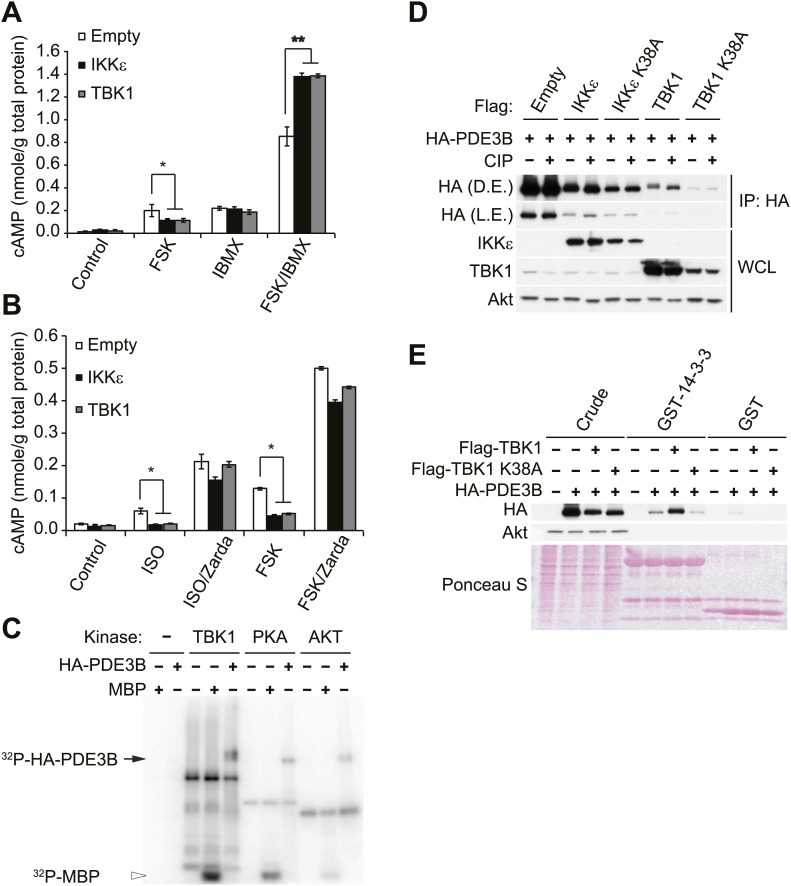
Figure 3. IKKε and TBK1 reduce cAMP levels through activation of PDE3B.

(A) cAMP levels from 3T3-L1 adipocytes expressing empty vector, Flag-IKKε, or Flag-TBK1 treated with or without 50 μM FSK, 250 μM IBMX, or together for 15 min. *p<0.05 and **p<0.0001. Performed in duplicate. (B) cAMP levels from 3T3-L1 adipocytes expressing empty vector, Flag-IKKε, or Flag-TBK1 treated with or without 10 μM ISO or 50 μM FSK together with or without 10 μM Zardaverine (Zarda) for 15 min. *p<0.05. Performed in duplicate. (C) 32P phospho-image of in vitro kinase reaction using either immunoprecipitated HA-PDE3B from HEK293T cells or 1 μg MBP (myelin basic protein) as a substrate with recombinant kinases as indicated. Results were replicated in multiple experiments. (D) Immunoblots of immunoprecipitation with anti-HA antibodies followed by treatment with or without CIP (top panel) and whole cell lysates (bottom panel) from Cos-1 cells co-expressing HA-PDE3B with Flag-IKKε/TBK1 or Flag-IKKε/TBK1 K38A. D.E. stands for dark exposure and L.E. stands for light exposure. Results were replicated in multiple experiments. (E) Immunoblots of GST-14-3-3 pulldown from HEK293T cells co-expressing HA-PDE3B with Flag-TBK1 or Flag-TBK1 K38A. Ponceau S staining shows the amount of beads used in GST-14-3-3 pulldown. Results were replicated in multiple experiments.

DOI: http://dx.doi.org/10.7554/eLife.01119.007

Figure 3.

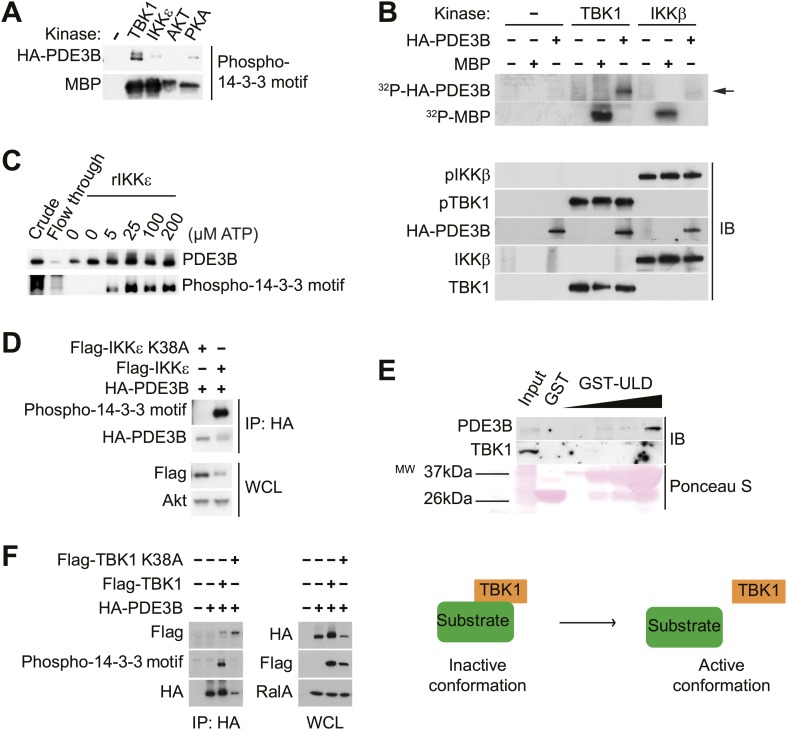
Figure 3—figure supplement 1. IKKε and TBK1 interact with PDE3B in a manner dependent on the activity of IKKε and TBK1.

Figure 3—figure supplement 1.

(A) Immunoblots of in vitro kinase reaction using either immunoprecipitated HA-PDE3B from HEK293T cells or 1 μg MBP (myelin basic protein) as a substrate with recombinant kinases as indicated. Results were replicated in multiple experiments. (B) Top panel: 32P phospho-image of in vitro kinase reaction using either immunoprecipitated HA-PDE3B from HEK293T cells or 1 μg MBP as a substrate with recombinant kinases as indicated. Bottom panel: Immunoblots (IB) of in vitro kinase reaction. Results were replicated in multiple experiments. (C) Immunoblots of in vitro kinase reaction using immunoprecipitated HA-PDE3B from HEK293T cells as a substrate with recombinant MBP-IKKε kinase (rIKKε) using increasing amounts of ATP as indicated. Lane 1: Whole cell lysates from HEK293T cells expressing HA-PDE3B, Lane 2: IP flow-through, Lane 3: IP without rIKKε. Results were replicated in multiple experiments. (D) Immunoblots of immunoprecipitation with anti-HA antibodies (top panel) and whole cell lysates (WCL, bottom panel) from HEK293T cells co-expressing HA-PDE3B with Flag-IKKε or Flag-IKKε K38A. Results were replicated in multiple experiments. (E) Immunoblots of GST-TBK1 ULD pulldown from 3T3-L1 adipocytes. Ponceau S staining shows the amount of beads used in GST-TBK1 ULD pulldown. Results were replicated in multiple experiments. (F) Immunoblots of immunoprecipitation with anti-HA antibodies (left panel) and whole cell lysates (right panel) from HEK293T cells co-expressing HA-PDE3B with Flag-TBK1 or Flag-TBK1 K38A. Results were replicated in multiple experiments. The schematic model suggests that TBK1 associates with its substrates in an inactive conformation and subsequently dissociate upon phosphorylation in an active conformation.