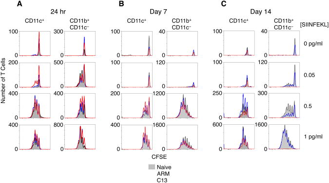
Figure 2. see also Figure S2: CD8+ T Cell stimulatory capacity of APCs from LCMV infected mice.
(A) Total proliferating CFSE-labeled OT-I T cells following culture with CD11c+ DC or CD11c− CD11b+ myeloid cells 24hr p.i. Proliferation was measured by flow cytometry after 3 days of culture with different amounts of SIINFEKL peptide.
(B) Total DC and myeloid cells from day 7 p.i. or day 14 p.i. (C) were bead purified and cultured with purified OVA-specific OT1 T cells as in previous figure.
Data are representative of 3 (A) or 2 (B-C) experiments.

