
Figure 2.
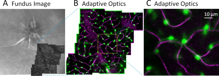
Simultaneous imaging of vascular perfusion and pericytes in vivo. (A) Wide field HRA Spectralis image shows approximately 30° field of mouse retina. Superimposed (dark) are motion-contrast AOSLO fields revealing capillary perfusion with micron-level resolution. (B) In vivo, two-channel imaging using AOSLO. Channel 1 collects NIR motion contrast that reveals capillary perfusion (magenta, moving blood cells). Channel 2 simultaneously images DsRed fluorescence (pericytes, green). (C) 5° AOSLO field shows the association of pericytes with perfused capillaries. Montage is from a single capillary stratification.