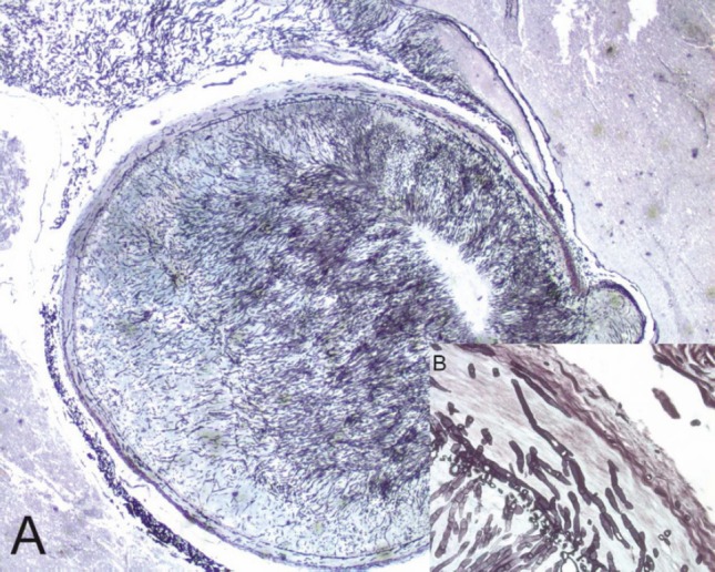
Figure 2. A mycotic thrombus occluding the lumen of the basilar artery (A) (2x original magnification, Grocott staining). The fungi showed typical broad, haphazardly branched hyphae that performed a basilary artery wall, inset (B) (40x original magnification, Grocott staining).

