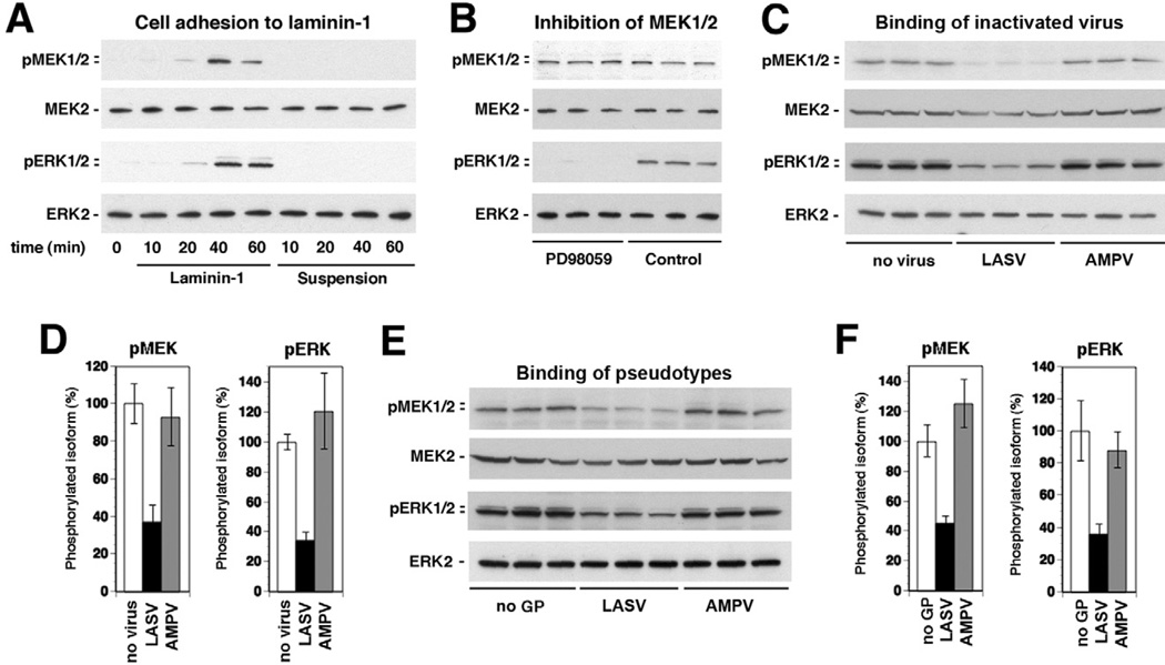
Fig. 4. Binding of inactivated LASV to cells perturbs laminin-induced activation of the ERK-MAP kinase pathway.
A. Phosphorylation of MEK and ERK in WI-26 VA4 cells in response to cell adhesion to laminin-1. Serum-starved cells were detached and seeded onto wells coated with laminin-1 or kept in suspension. At the indicated time points, total-cell lysates were prepared and subsequently analysed for the presence of MEK1/2 and ERK1/2 as well as the phosphorylated forms of the kinases using specific antibodies.
B. ERK is phosphorylated via MEK: suspensions of serum-starved cells were incubated with the MEK-specific inhibitor PD98059 (50 µM) or solvent control for 30 min and then plated onto wells coated with laminin-1 for 40 min. The phosphorylated forms of MEK1/2 and ERK1/2, MEK2 and ERK2 were detected in total-cell lysates.
C. Binding of inactivated LASV to cells reduces laminin-induced activation of MEK and ERK: serum-starved WI-26 VA4 cells were detached, mixed with inactivated LASV or AMPV (10 particles per cell), or no virus, and plated onto wells coated with laminin-1. After 40 min of cell adhesion, total-cell lysates were prepared and analysed for the presence of phosphorylated MEK and ERK as in (A).
D. Quantification of (C): the signal for phosphorylated MEK1/2 and ERK1/2 in the control samples set as 100% (n = 3, ±SD).
E and F. Binding of LASV pseudotypes reduces laminin-induced activation of MEK and ERK: experiment was performed as in (C) and (D) using retroviral pseudotypes of LASV and AMPV (10 PFU per cell) and pseudotypes without GP (no GP).

