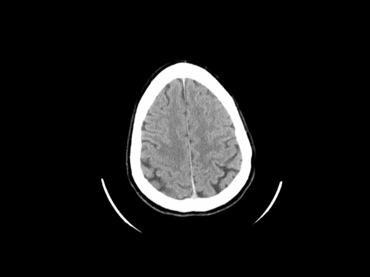
Figure 1.
Head Computerized Tomography (CT) Without Contrasta
aHead CT obtained in the course of the emergency room evaluation. This brain scan was read to be within normal limits. In retrospect, one can use the T2-weighted magnetic resonance imaging scan to guide the search and identify an area of very mild hypoattenuation in the right frontal lobe corresponding to the tumor.

