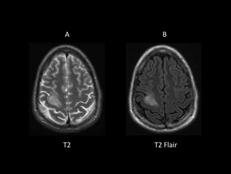
Figure 3.
T2 (A) and T2-FLAIR (B) Magnetic Resonance Image Sequencesa
aThis pulse sequence clearly identifies the right frontal tumor as a hyperintense lesion in both the T2 (A) and T2-FLAIR (B). Notice the contrast difference between the 2 images: once the fluid hyperintense signal is suppressed with FLAIR, the lesion increases its contrast.
Abbreviation: FLAIR = fluid attenuated inverted recovery.

