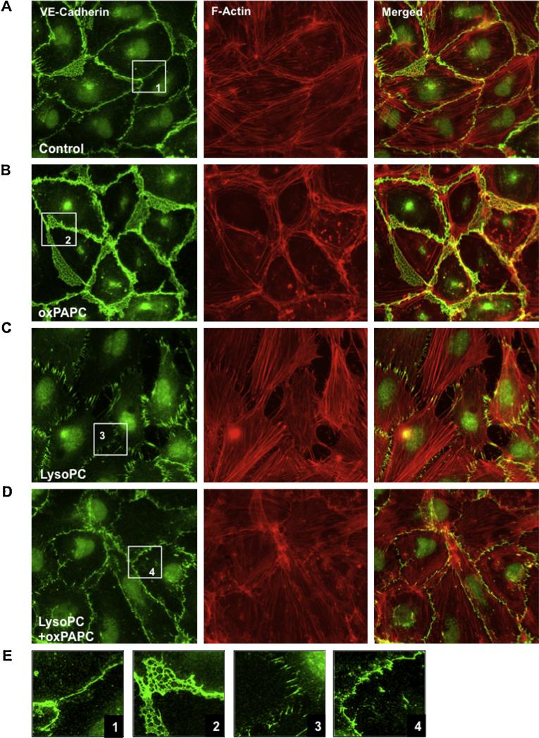
Fig. 8.
Effect of lyso-PC, oxPAPC and their combination on pulmonary endothelial cell cytoskeleton and monolayer integrity. Human pulmonary EC monolayers were left untreated (A); stimulated for 30 min with 20 μg/ml oxPAPC (B); 10 μg/ml lysoPC (C); or cotreated with 10 μg/ml lysoPC and 20 μg/ml oxPAPC (D). Analysis of actin cytoskeletal remodeling was performed by immunofluorescent staining with Texas Red phalloidin (red). Adherens junctions were detected by staining with VE-cadherin (green). Right panels show merged images of F-actin and VE-cadherin staining. Insets (E) depict higher magnification images with details of actin and adherens junction structures in control, lysoPC and oxPAPC stimulated cells. (For interpretation of the references to color in this figure legend, the reader is referred to the web version of the article.)

