
Figure 11.
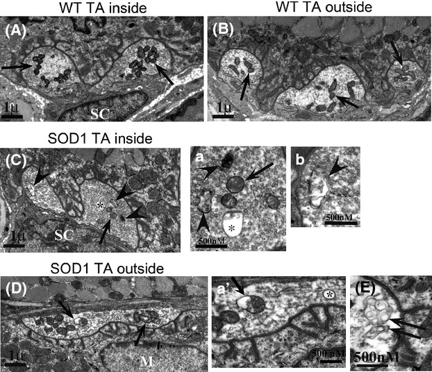
(A and B) Normal NMJ appearance in the inner TA (types IIa and IIb) and outer TA (primarily type IIb), respectively in P30 animals. Arrows point to normal compact terminal mitochondria. (C) In SOD1 animals, alterations in inner TA, enlarged in a and b, while (D) shows outer TA, enlarged in a'. Asterisks in a and a' are inside of large vacuoles (>100 nm diameter), a common feature in SOD1 animals. Arrows point out swollen mitochondria, and arrowheads point out abnormalities that appear autophagic-like. Double arrows in (E) point to abnormal whorls in a terminal from the outside portion of the TA. Overall, abnormalities of all types were seen 2–3 times more frequently in SOD1 than WT. Representative images are shown. Five animals of each genotype were examined.