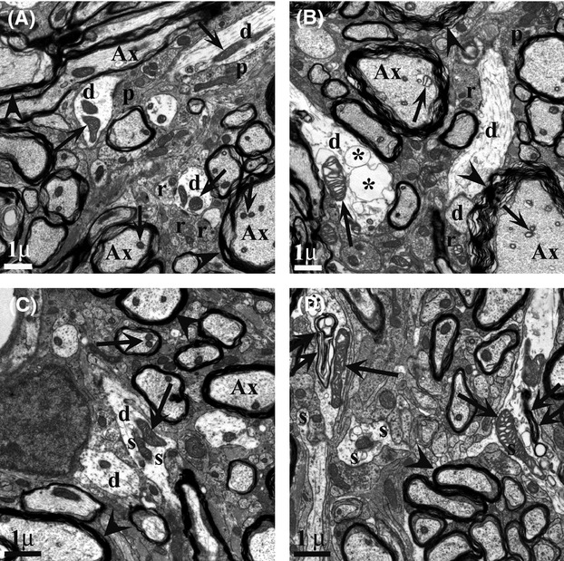
Figure 17.

Distal dendrites in SOD1 MNs exhibit vacuolated mitochondria and large cytoplasmic vacuoles, and axons have fragmented myelin sheaths. Synapse types on MN distal dendrites (d) at P30 in white matter adjacent to VH showed a decrease in type I (r) synapses similar to that seen for MNs, with no significant decrease in type II (p). Note distension of mitochondrial cristae (arrows) in SOD1 (B) compared with WT (A) in both axons (Ax) and dendrites, as well as poor condition of myelin sheath (arrowheads) and presence of large vacuoles (*) in SOD1 (n = 5 animals each, WT and SOD1). Double arrows in D point to large autophagic-like bodies in dendrites. For distal dendrites, using the same levels as used for MN soma evaluation in Figure 16, 20–30 dendrites with synapses from each animal used above were located in the ventro-lateral white matter and photographed at 16,000×. Only synapses with a clear synaptic density and presynaptic vesicles were scored.
